-
Call Us044-42123262
-
Email Us sparksdentalcentre@gmail.com
General dentistry
Maxillofacial trauma
Anatomy The treatment of maxillofacial trauma is quite complex and is best explained through the anatomy of the maxillofacial region. The region is divided into 3 parts: the upper face constituting of the frontal bone and the frontal sinus, the mid-face which constitutes of the nasal, ethmoid, zygomatic and maxillary bones and the lower face containing the mandible.The trigeminal nerve is responsible to deliver sensations to the skin on the face, besides motor functions such as chewing and biting.


Types of trauma
Frontal bone fractures happen when there is a blow to the forehead involving the frontal sinus also.
Orbital floor fracture is an isolated fracture involving the medial wall.
Nasal fractures occur when there is direct trauma to the nasal area.
NOE or nasoethmoidal fractures travel from the ethmoid bone to the nose with resultant damage to the lacrimal apparatus, the nasofrontal duct or the medial canthus.
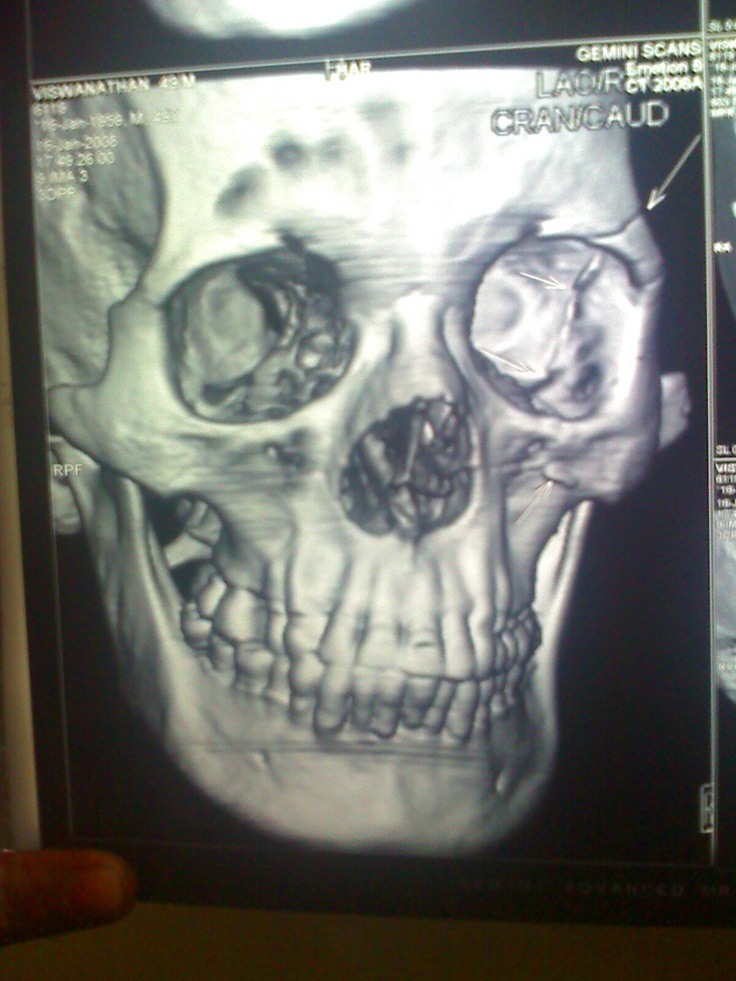
ZMC or zygomaticomaxillary complex fractures are also as a result of direct trauma and affect the orbital floor and the infraorbital foramen and extending into the zygomaticomaxillary, zygomaticofrontal and zygomaticotemporal sutures.
Zygomatic arch fractures occur when there is a direct trauma induced to the zygomatic arch and can involve the zygomaticotemporal suture.
Maxillary fractures are of three types Le Fort I, II or III which is the horizontal maxillary fracture, a pyramidal fracture and craniofacial dysfunction respectively.
Alveolar fractures involve the alveolar area of the mandible or maxilla which usually occurs due to direct but low-energy impacts.
Mandibular fractures are secondary to the U-shape of the jaw, occurring in multiple locations.
Panfacial fractures involve the upper face, mid face and lower face combining 3 to 4 facial units.
Diagnosis and investigation
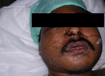
A physical examination is conducted to reveal if there is bleeding from the nose, nasal blockage or skin lacerations. There could be bruising surrounding the eyes and the distance between the eyes could be widened, with changes in vision and movement of the eyes.
Fractures are investigated with appropriate x-rays and CT scan of the head, upper face, mid face and lower face are conducted depending on the nature of the trauma.
A CBC is done to check for haemoglobin and haematocrit if excessive bleeding is noted. SMA-20 and bhCG studies are also conducted.
TreatmentUsually a surgical intervention is the treatment option if any of the injuries prevent normal functioning of the organs. The aim of the treatment is to clear the airway, control the bleeding, fix the bone segments by treating the fracture and even try and prevent scarring.


